Pelvic Anatomy Bones / Pelvis Gender Differences Of Pelvic Anatomy / Some pelvic fractures involve breaking more than one of the bones, and these are particularly serious as the bones are more likely to slip out of line.
Pelvic Anatomy Bones / Pelvis Gender Differences Of Pelvic Anatomy / Some pelvic fractures involve breaking more than one of the bones, and these are particularly serious as the bones are more likely to slip out of line.. The pelvis is a ring structure made up of three bones: The outlet is formed by the pubic arch, ischial spines, sacrotuberous ligaments, and the coccyx. The pelvis is composed of the two pelvic bones and the sacrum and coccyx (the pelvic bones are also known as the coxal, innominate, or hip bones) (fig. This is a secondary cartilaginous joint of pelvis between the bodies of the right and left pubic bones. This quiz is unlabeled so it will test your knowledge on how to identify these structural locations (iliac crest, ischial spine, acetabulum, superior ramus of pubis, posterior superior/inferior iliac spine, lessier.
The pelvic bones and the sacrum. The pelvic girdle (hip girdle) is formed by a single bone, the hip bone or coxal bone (coxal = hip), which serves as the attachment point for each lower limb. The femur is the largest bone in the body and the only bone of the thigh. A pelvic fracture is a break in any one of those bones. When the two hip bones are combined with the sacrum and coccyx of the axial skeleton, they are referred to as the pelvis.the right and left hip bones also converge anteriorly to attach to each other at the pubic symphysis (figure 8.3.1).
Clinical Anatomy Of The Vulva Vagina Lower Pelvis And Perineum Glowm from resources.ama.uk.com When you are taking anatomy and physiology you will be required to know the anatomical structure locations of the pelvis. List the arterial & nerve supply list the lymph & venous drainage of the pelvis. Formed by the left and right hip bones, the pelvic girdle connects the lower limb (leg) bones to the axial skeleton. Über 7 millionen englischsprachige bücher. These bones connect the axial skeleton to the lower limbs, and therefore play a role in bearing the weight of the upper body. When the two hip bones are combined with the sacrum and coccyx of the axial skeleton, they are referred to as the pelvis.the right and left hip bones also converge anteriorly to attach to each other at the pubic symphysis (figure 8.3.1). The inlet to the pelvic canal is at the level of the sacral promontory and superior aspect of the pubic bones. Each pelvic bone (hip bone) is made by the combination three bones namely, the ilium, pubis, and ischium.
The pelvic skeleton is formed posteriorly (in the area of the back), by the sacrum and the coccyx and laterally and anteriorly (forward and to the sides), by a pair of hip bones.
Formed by the left and right hip bones, the pelvic girdle connects the lower limb (leg) bones to the axial skeleton. These bones connect the axial skeleton to the lower limbs, and therefore play a role in bearing the weight of the upper body. The pelvis is a ring structure made up of three bones: They meet at the triradiate cartilage, which fuses by the age of 16 years. A 3d rotatable model of the bony structures of the pelvis: The pelvic bones and the sacrum. Describe the anatomy of the pelvic wall, bones, joints & muscles. This quiz is unlabeled so it will test your knowledge on how to identify these structural locations (iliac crest, ischial spine, acetabulum, superior ramus of pubis, posterior superior/inferior iliac spine, lessier. This is a secondary cartilaginous joint of pelvis between the bodies of the right and left pubic bones. The counterrotation of the bones continues all down the leg and through the foot. Some pelvic fractures involve breaking more than one of the bones, and these are particularly serious as the bones are more likely to slip out of line. The pelvic skeleton is formed posteriorly (in the area of the back), by the sacrum and the coccyx and laterally and anteriorly (forward and to the sides), by a pair of hip bones. The ilium, pubis and ischium.
A pelvic fracture is a break in any one of those bones. There are two hip bones, one on the left side of the body and the other on the right. List the arterial & nerve supply list the lymph & venous drainage of the pelvis. Über 7 millionen englischsprachige bücher. The range of motion is increased temporarily in pregnancy in which all the ligaments joints of the pelvis become loose, under the influence of hormones, to facilitate the delivery of the fetus.
Clinical Anatomy Of The Vulva Vagina Lower Pelvis And Perineum Glowm from resources.ama.uk.com This is a secondary cartilaginous joint of pelvis between the bodies of the right and left pubic bones. The pelvic spine is the posterior portion of the pelvis below the lumbar spine, composed of the sacrum and coccyx. Formed by the left and right hip bones, the pelvic girdle connects the lower limb (leg) bones to the axial skeleton. The low back is defined by the lumbar spine, and the pelvis is defined by the bones of the pelvic girdle. The pelvic fl oor constitutes the caudal border of the human's visceral cavity. The sacrum and the two innominate bones. 10+ bones of pelvis anatomy 11 photos of the 10+ bones of pelvis anatomy hip anatomy bones, pelvic bone anatomy, pelvis muscle anatomy, pelvis skeletal anatomy, radius bone anatomy, three bones of the pelvis, tibia bone anatomy, human anatomy, hip anatomy bones, pelvic bone anatomy, pelvis muscle anatomy, pelvis skeletal anatomy, radius bone. Each hip bone contains three bones — the ilium, ischium, and pubis — that fuse together as we grow older.
Some pelvic fractures involve breaking more than one of the bones, and these are particularly serious as the bones are more likely to slip out of line.
Together, they form the part of the pelvis called the pelvic girdle. The pelvic girdle, also known as the hip bone, is composed of three fused bones: The sacrum and the two innominate bones. The femur is the largest bone in the body and the only bone of the thigh. Describe the components & function of the pelvic diaphragm. Formed by the left and right hip bones, the pelvic girdle connects the lower limb (leg) bones to the axial skeleton. A full understanding of pelvic anatomy is required to treat pelvic fractures, to prevent iatrogenic injuries, and to provide the best results. They meet at the triradiate cartilage, which fuses by the age of 16 years. Über 7 millionen englischsprachige bücher. Each hip bone, in turn, is firmly joined to the axial skeleton via its attachment to the sacrum of the vertebral column. The pelvic bones in a male are smaller and narrower than in females, which is larger and wider. During childhood, these sections are separate bones, joined by the triradiate cartilage. Each hip bone contains three bones — the ilium, ischium, and pubis — that fuse together as we grow older.
Each hip bone consists of 3 sections, ilium, ischium, and pubis. These bones also act as attachments for many muscles and ligaments within the pelvis and lower limbs. 5 out of 5 stars (1,608) $ 12.95. The pelvic bones in a male are smaller and narrower than in females, which is larger and wider. The hip bone has three parts:
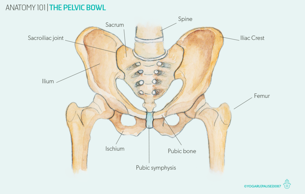
Anatomy 101 The Pelvic Bowl Yogaru from images.squarespace-cdn.com The pelvis is a ring of bone at hip level, made up of several separate bones. Differentiate the different types of the female pelvis. A full understanding of pelvic anatomy is required to treat pelvic fractures, to prevent iatrogenic injuries, and to provide the best results. Learning pelvic anatomy is composed of learning bones, muscles, ligaments, nerves and vascular supply. The ilium, ischium and the pubic bone. In the franklin method we call the natural and safe way bones move the bone rhythms (figure 23). When the two hip bones are combined with the sacrum and coccyx of the axial skeleton, they are referred to as the pelvis.the right and left hip bones also converge anteriorly to attach to each other at the pubic symphysis (figure 8.3.1). The pelvic skeleton is formed posteriorly (in the area of the back), by the sacrum and the coccyx and laterally and anteriorly (forward and to the sides), by a pair of hip bones.
Differentiate the different types of the female pelvis.
Pelvis keychain, human anatomy pelvic bone keyring in pewter farjil. The pelvic bones and the sacrum. This video provides an overview of the bones of the pelvis narrated by one of our awesome anatomists using the latest 3d anatomy software. There are four joints within the pelvis: The bones of the pelvis are the hip bones, sacrum, and coccyx. Formed by the left and right hip bones, the pelvic girdle connects the lower limb (leg) bones to the axial skeleton. 5 out of 5 stars (1,608) $ 12.95. Each hip bone, in turn, is firmly joined to the axial skeleton via its attachment to the sacrum of the vertebral column. Each hip bone contains three bones — the ilium, ischium, and pubis — that fuse together as we grow older. The pelvis is a ring of bone at hip level, made up of several separate bones. The hip bones join to the upper part of. Describe the components & function of the pelvic diaphragm. The pelvis consists of two innominate bones and the sacrum to which coccyx is attached.
List the arterial & nerve supply list the lymph & venous drainage of the pelvis pelvic anatomy. The pelvis is a ring of bone at hip level, made up of several separate bones.


0 Komentar